
Varicose vein
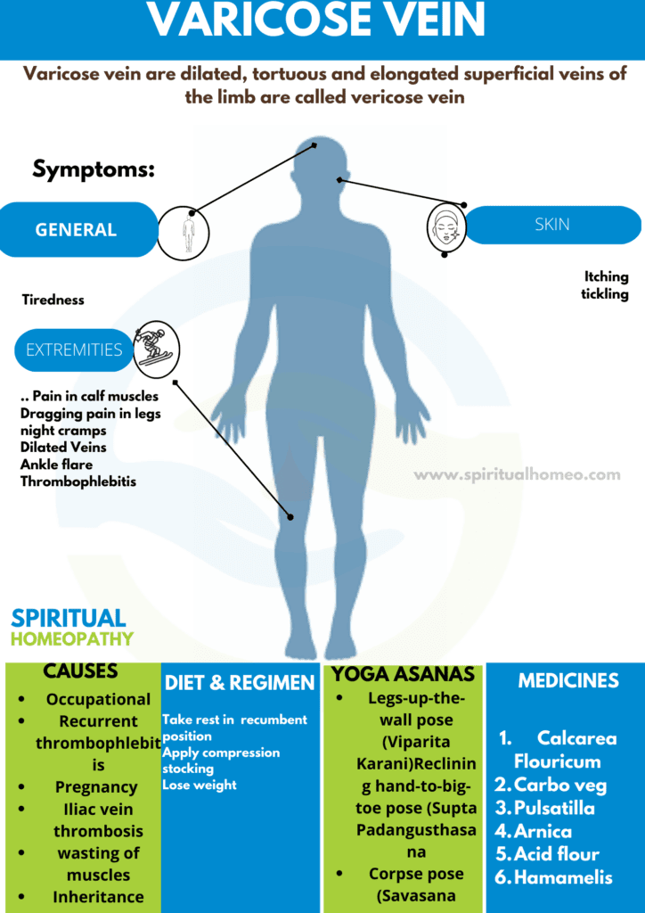
Varicose vein are dilated, tortuous and elongated superficial veins of the limb.
- Varicosity is the penalty for verticality against gravity.
- The blood has to flow from the lower limbs into the heart against gravity because of the upright posture of human beings.
- In many cases, varicose veins are asymptomatic.
- Raised intra-abdominal pressure also precipitates varicose veins, more commonly in females due to repeated pregnancy.
- The complications of varicose veins are responsible for hospitalization of the patient.
Varicosities are more common in lower limb because of erect posture and long column of blood has to be supported which can lead to weakness and incompetency of valves.
1.Primary varicosities due to i.e.:
- Congenital either incompetence or absence of valves.
- Weakness or wasting of muscles: defective connective tissue also smooth muscle in the venous wall.
- Stretching of deep fascia.
- Inheritance (family history) with FOXC2 gene.
- Klippel – Trenaunay syndrome.
2. Secondary varicosities i.e.:
- Recurrent thrombophlebitis,
- Occupational – standing for long hours (e.g. traffic police, guards).
- Obstruction to venous return like abdominal tumours, retroperitoneal fibrosis, lymphadenopathy.
- Pregnancy (due to progesterone hormone).
- AV malformations: Either Congenital or acquired.
- Iliac vein thrombosis.
- Height: Tall individuals suffer more
- Weight: Obesity may weaken vein wall
- Occupation: e.g. Hotel workers, police officers, shopkeepers, tailors
- Side: Left is comparatively affected more than the right
- Age and sex: Not very clear.
- Generally, Incompetence of venous valves causes stasis of blood.
- Furthermore, Chronic ambulatory venous hypertension causes defective microcirculation.
- So, RBC diffuses into tissue planes also lysis of RBC’s start.
- This leads to release of haemosiderin.
- It leads to Pigmentation, Dermatitis also Capillary endothelial
- Results in to Prevention of diffusion also exchange of nutrients causes severe
- Lastly, It leads to chronic venous ulceration.
- Long saphenous vein varicosity.
- Short saphenous vein varicosity.
- Varicose veins due to perforator incompetence.
- Thread veins (or dermal flares/telangiectasias/spider
- veins are 0.5 – 1 mm in size): Are small varices in the skin usually around ankle which look like dilated,
- Red or purple network of veins.
- Reticular varices (1-3 mm in size): Are slightly larger
- Varices than thread veins located in subcutaneous region.
- Combinations of any of above.
- Majority of the patients present with dilated veins in the leg.
- They are minimal to start with and at the end of the day they are sufficiently large because of the venous engorgement.
- Dragging pain in the leg or dull ache is due to heaviness.
- Night cramps occur due to change in the diameter of veins.
- Aching pain is relieved at night on taking rest or elevation of limbs.
- Sudden pain in the calf region with fever and Oedema on the ankle region suggests deep vein thrombosis.
- Patients can present with ulceration, eczema , dermatitis and bleeding.
- Symptoms of pruritus/itching and skin thickening.
Signs:
Inspection (should be done in standing position)
- Dilated veins are present in the medial aspect of leg and the knee. Sometimes they are visible in the thigh also.
- Single dilated varix at SF junction is called saphenous varix. It is due to saccular dilatation of the upper end of long saphenous vein at the saphenous opening.
- Veins are tortuous and dilated.
- A localized, dilated segment of the vein, if present, is an indication of a blowout. It signifies underlying perforator.
- Ankle flare is a group of veins near the medial malleolus.
- Complications such as ulceration, bleeding, eczema and dermatitis may be present.
- Healed scar indicates previous ulceration.
- Look at the popliteal fossa region also.
Palpation
- First palpate along the whole length of vein. Look for tenderness. If present, it indicates thrombophlebitis.
- Vein which is thrombosed will feel as a firm/hard nodule.
-
Cough impulse test (Morrissey’s test):
- This test should be done in the standing position.
- The examiner keeps the finger at SF junction and asks the patient to cough.
- Fluid thrill, an impulse felt by the fingers, is indicative of ‘saphenofemoral incompetence’.
-
Trendelenburg test:
Method:
- The patient is asked to lie on the couch in the supine position.
- The leg is elevated above the level of heart and the vein emptied.
- SF junction is occluded with the help of the thumb (or a tourniquet) and the patient is asked to stand.
Trendelenburg I i.e.:
- Release the thumb or tourniquet immediately.
- Rapid gush of blood from above downwards indicates saphenofemoral incompetence.
Trendelenburg II i.e.:
- The pressure at the SF junction is maintained without releasing the thumb or tourniquet.
- The patient is then asked to stand.
- Slow filling of the long saphenous is seen. It is due to per/orator incompetence (retrograde flow of blood).
-
Multiple Tourniquet test:
- It is done to find out exact site of perforators
Method i.e.:
- The patient is asked to lie supine on the couch.
- The vein is emptied by elevation.
- As the name suggest 3-5 tourniquets (multiple) can be applied.
- However, more tourniquets are applied, the exact localization c the perforators can be made out but it is not practical.
- There are mainly ankle, knee and thigh perforatons.
- Hence, four tourniquets can be applied at various level as mentioned below.
- 1st Tourniquet: At the level of saphenofemoral junction (SF junction).
- 2nd Tourniquet: At the level of middle of the thigh, t, occlude perforator in the Hunter’s canal.
- 3rd Tourniquet: Just below the knee.
- 4th Tourniquet: Palm breadth (lower third of the leg above medial malleolus/ankle.
- Ask the patient to stand and observe appearance of veins
Inference i.e.:
- Appearance of veins between first and second tourniquets indicates incompetence of thigh perforators between second and third indicates incompetence of knee, perforators and below the fourth tourniquet indicates incompetence of ankle perforators.
- Most commonly, below knee and ankle perforators are incompetent.
- On releasing the tourniquets one by one from be upwards, sudden retrograde filling of the veins occurs.
-
Schwartz test:
- It is done with the patient in the standing position.
- Place the fingers of the left hand over a dilated segment of the vein and with the right index finger tap the vein below.
- A palpable impulse suggests a superficial column of blood in the vein and it also suggests incompetence of the valves in between the segment of the vein.
-
Modified Perthes’ test:
- It done to rule out deep vein thrombosis.
- The patient ask to stand, the tourniquet is applied at SF junction and he ask to have a brisk walk.
Inference i.e.:
- If the patient complains of severe pain in calf region or if superficial veins become more prominent, it is an indication of deep vein thrombosis and is a contraindication for surgery.
-
Fegan’s method (test):
- It done to detect the site of perforators.
- The patient ask to stand. The varicosity is marked with methylene blue and he is asked to lie down.
- The leg elevate to empty the vein and the vein palpate throughout its course.
- The defects in the deep fascia have a circular, buttonhole consistency.
Hand held Doppler i.e.:
- It is the first, mm1mum level investigation to do before treating a patient with venous disease.
- When the blood flows, the wave emits a signal – the Doppler signal.
- If there is SF incompetence, fo1ward and backward flow can detect.
- Biphasic signal: Gently squeeze calf muscles and the flow can assess by Doppler probe in the SF junction.
- It can also pick up accessory long saphenous vein in the groin.
- It is not an investigation to identify the perforators.
Duplex ultrasound imaging i.e.:
- In this investigation high resolution B-mode ultrasound imaging and Doppler ultrasound use.
- It helps in getting images of veins, measure flow in all lower limb veins.
- Origin of venous ulcers and varicose veins can also assess.
Venography i.e.:
- Both ascending and descending venography can done in a case of deep vein thrombosis.
- It is an invasive procedure and risk of spreading infection and septicemia is present.
- Duplex ultrasonography has largely replaced this investigation (rarely done now).
- Varicography refers to injecting contrast into surface veins (indicated in recurrent varicose veins).
- It can follow by sclerosant injections.
Plethysmography i.e.:
- They base on measurement of volume changes in the leg.
- By placing a light-emitting diode above medial malleolus and patient performing tiptoe movements, venous recovery time can measure.
- This call phot plethysmography (PPT).
- Lymphoedema
- A-V malformation
- Orthostatic Oedema
- Renal and cardiac disease
- Hepatic causes
- Vasculitis
- Metabolic diseases like gout, myxedema, and morbid obesity
- Chronic infections like tuberculosis, syphilis.
- Patients with normal deep veins should offer treatment to obliterate any sites of saphenous or perforator reflux.
- Elastic stockings should prescribe for all patients with evidence of post-thrombotic deep vein damage and these remain an alternative treatment for patients with superficial venous disease who decline intervention.
Homeopathy treats the person as a whole. It means that homeopathic treatment focuses on the patient as a person, as well as his pathological condition. The homeopathic medicines selected after a full individualizing examination and case-analysis.
which includes
- The medical history of the patient,
- Physical and mental constitution,
- Family history,
- Presenting symptoms,
- Underlying pathology,
- Possible causative factors etc.
A miasmatic tendency (predisposition/susceptibility) also often taken into account for the treatment of chronic conditions.
What Homoeopathic doctors do?
A homeopathy doctor tries to treat more than just the presenting symptoms. The focus is usually on what caused the disease condition? Why ‘this patient’ is sick ‘this way’?.
The disease diagnosis is important but in homeopathy, the cause of disease not just probed to the level of bacteria and viruses. Other factors like mental, emotional and physical stress that could predispose a person to illness also looked for. No a days, even modern medicine also considers a large number of diseases as psychosomatic. The correct homeopathy remedy tries to correct this disease predisposition.
The focus is not on curing the disease but to cure the person who is sick, to restore the health. If a disease pathology not very advanced, homeopathy remedies do give a hope for cure but even in incurable cases, the quality of life can greatly improved with homeopathic medicines.
Homeopathic Medicines for Varicose vein :
The homeopathic remedies (medicines) given below indicate the therapeutic affinity but this is not a complete and definite guide to the homeopathy treatment of this condition. The symptoms listed against each homeopathic remedy may not be directly related to this disease because in homeopathy general symptoms and constitutional indications also taken into account for selecting a remedy, potency and repetition of dose by Homeopathic doctor.
So, here we describe homeopathic medicine only for reference and education purpose. Do not take medicines without consulting registered homeopathic doctor (BHMS or M.D. Homeopath).
Medicines:
-
Calcarea Flouricum
- This tissue salt relaxes the Elastic Fibers, especially of veins and glands.
- Glands enlarge also Become Stony Hard.
- Veins dilated also become varicose, inflamed.
- Weakness, in the morning.
- Besides this, Feeling of fatigue all day.
- Aggravation at beginning of motion, whereas Amelioration by continue motion.
-
Hamamelis Verginica:
- Venous congestion.
- Varicose veins with bruised soreness of affected parts.
- Furthermore, Acts upon the coats of veins causing relaxation with consequent engorgement.
- Tired feeling especially in legs.
- Chilliness in back also hips, extending down legs.
- Bleeding veins.
-
Pulsatilla:
- Drawing, tensive pains in thighs also legs, with restlessness, sleeplessness and chilliness.
- Pain in limbs, shifting rapidly.
- Tensive pain, letting up with a snap.
- Suffering worse specifically from letting the affected limb hang down.
- Legs feel heavy also weary.
- Lastly, Feet red, inflamed, swollen.
-
Carbo vegetablis:
- Disintegration also imperfect oxidation.
- Body becomes blue, icy-cold.
- Venous congestions. Heavy, stiff, feel paralyzed; limbs go to sleep; additionally want of muscular energy, joints weak.
- Cold from knees down.
- Toes red, swollen, burning pains in bones also limbs
- Cramps in soles, feet numb also sweaty.
-
Flouricum Acidum:
- Varicose veins.
- Distended blood vessels.
- Complaint of old age, feel weak.
- Ulcers; additionally red edges and vesicles.
-
Arnica :
- Generally, Limbs and body ache as if beaten; joints as if sprained.
- Relaxed blood vessels, black also blue spots.
- Tendency to tissue degeneration. sore, unimpressive, bruised feeling. Pain in back also limbs.
- Soreness after overexertion. Great fear of being either touched or approached.
- Tingling and itching which moves from place to place; In detail, after scratching,
- Besides this, Itching begins somewhere else.
- All in all, Blood vessels are relaxed, causing ecchymosis, blue-black spots; with tendency to hemorrhage; epistaxis.
- Consult best homeopathy doctors at spiritual homeopathy clinics Hyderabad. we also provide online consultation in homeopathy .
we are specialized for chronic ailments like thyroid ,diabetes, sexology problems, dermatology complaints, gastric complaints, rheumatoid and osteo arthritis, respiratory complaints, migraine and more. We have a team of expert doctors. WE are treating patients in more than 60 countries like India, Nepal, Bhutan, UAE, Saudi Arabia, Kuwait, Canada, England, Holland, China, Srilanka,Germany, France, USA,UK, Australia, New Zealand, Russia, Afghanistan,Myanmar and many other Asian and European countries. We have Expert Team -specialized in Homeopathy for kids, Homeopathy treatment for Children. - Book Your Video consultation or In clinic appointment now.
- Take rest with leg elevated above heart level.
- Apply compression stockings.
- Should lose weight.
What is Varicose vein ?
Varicose vein are dilated, tortuous and elongated superficial veins of the limb.
Homeopathic Medicines used by Homeopathic Doctors in treatment of Varicose vein ?
- Calcarea Flouricum
- Hamamelis Verginica
- Pulsatilla
- Carbo vegetablis
- Flouricum Acidum
- Arnica
What causes Varicose vein ?
- Congenital incompetence or absence of valves.
- Weakness or wasting of muscles
- Stretching of deep fascia.
- Inheritance (family history)
- Klippel – Trenaunay syndrome
- Recurrent thrombophlebitis,
- Occupational
What are the symptoms of Varicose vein ?
- Dilated veins in the leg
- Dragging pain in the leg or dull ache is due to heaviness.
- Night cramps occur due to change in the diameter of veins
- Aching pain is relieved at night on taking rest or elevation of limbs
- SRB’S manual of surgery 3rd edition
- Manipal manual of surgery 4th edition
- Bailey and Love short practice of surgery 25th edition
- Materia medica of homoeopathic medicine by DR. S.R.Phatak.
- Homoeopathic Body-System Prescribing – A Practical Workbook of Sector Remedies by Isaac Golden.